Clinical navigator
When the problem is neural,
localization changes everything.
Choose an area, then open the relevant condition. Each card gives a concise view of symptoms, assessment and treatment options—without inviting self-diagnosis.
Explore by region
Select a clinical area.
Brachial plexus and nerve transfers
The brachial plexus is the nerve network travelling from the neck to the shoulder, arm and hand. Injury may range from temporary stretch to rupture or root avulsion, making severity and timing central to reconstruction.
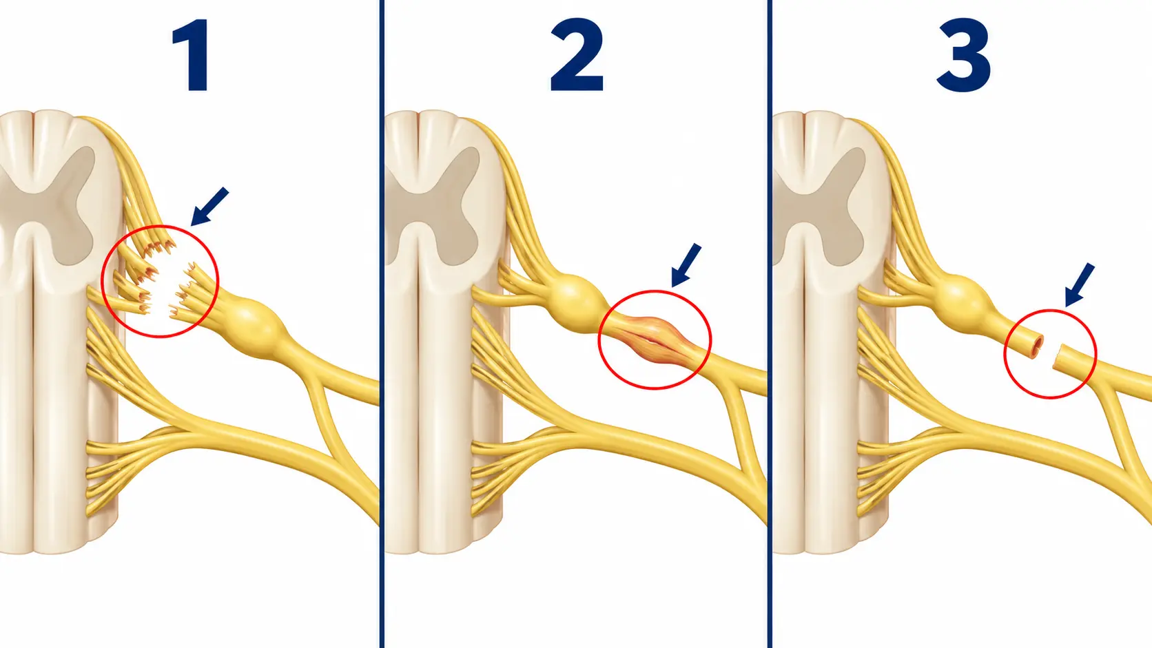
Brachial plexus injurySymptoms and assessment
Possible features include electric or burning pain, weakness or paralysis in part of the limb, sensory loss and sometimes severe neuropathic pain. The problem is usually unilateral.
How is it assessed?
Each movement and sensory territory is mapped, urgent vascular or spinal injury is excluded, and EMG/nerve-conduction studies and imaging are selected according to the mechanism. Serial examinations may show whether recovery has begun.
Repair, grafting and nerve transferFunctional reconstruction
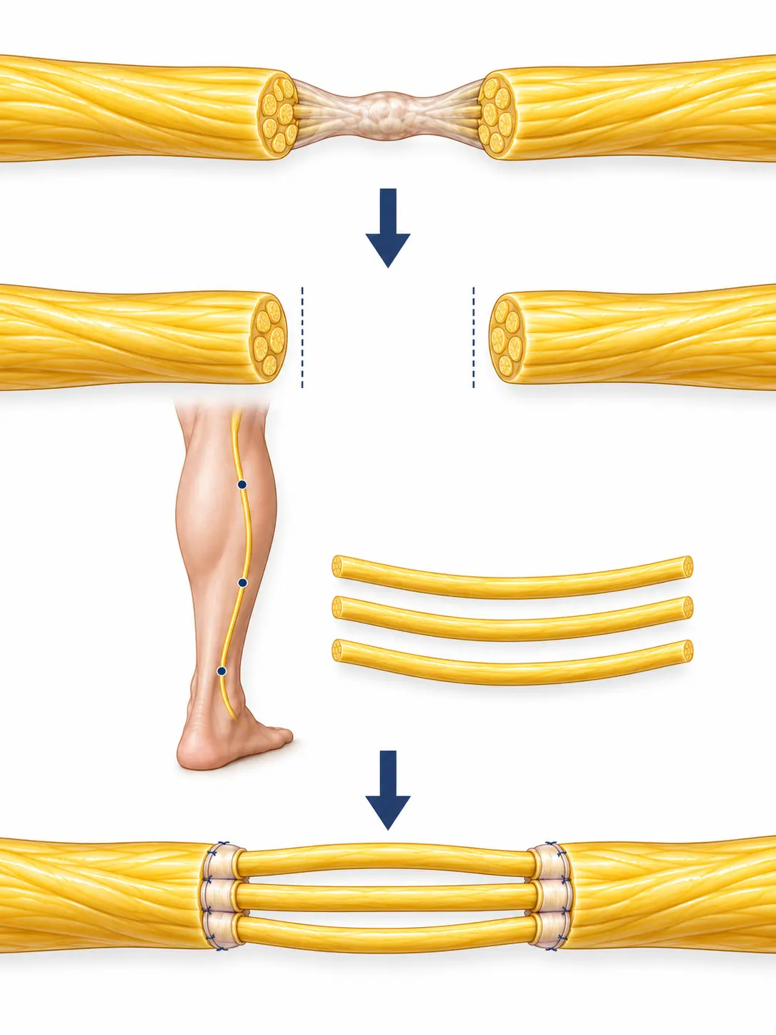
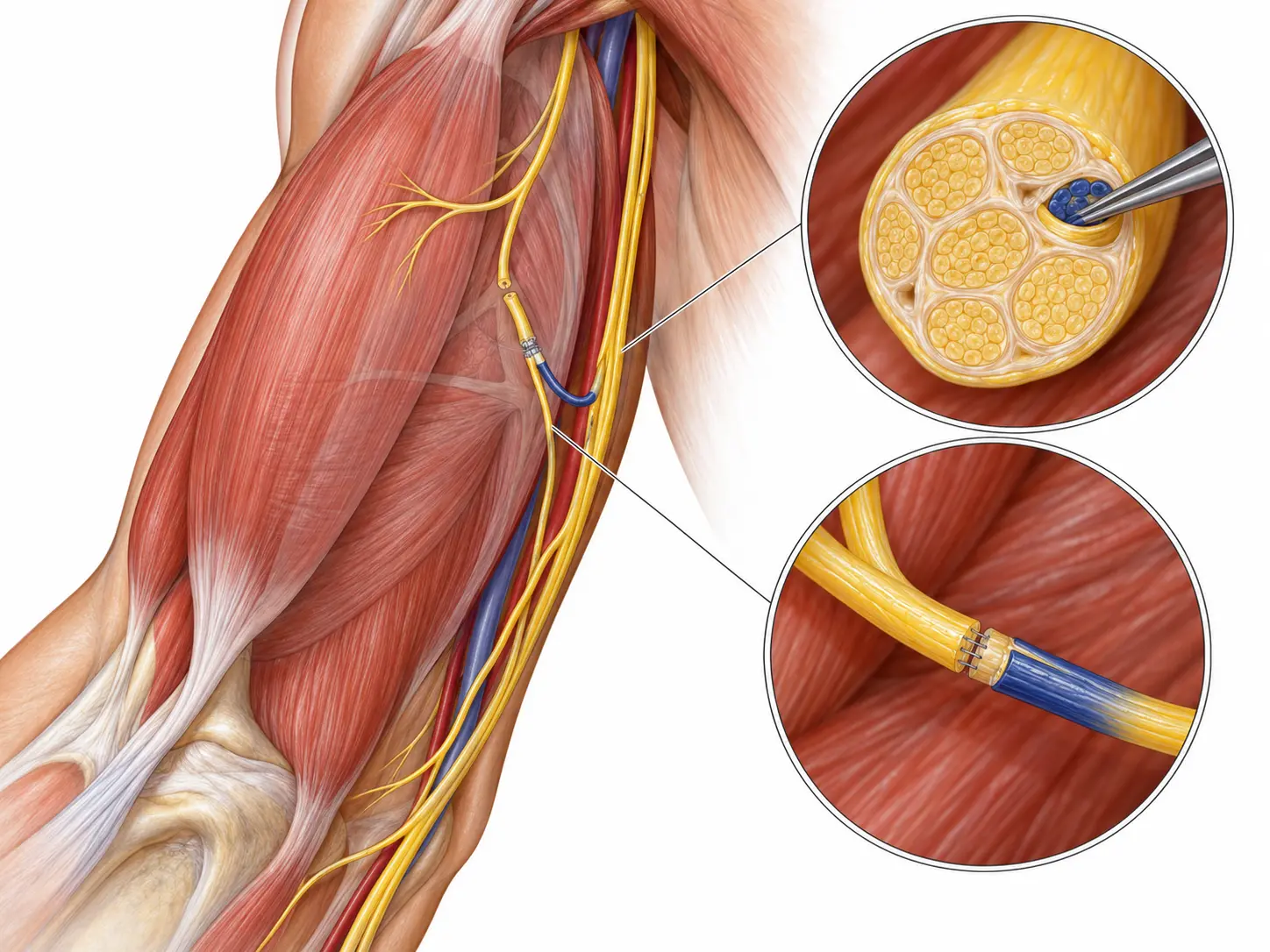
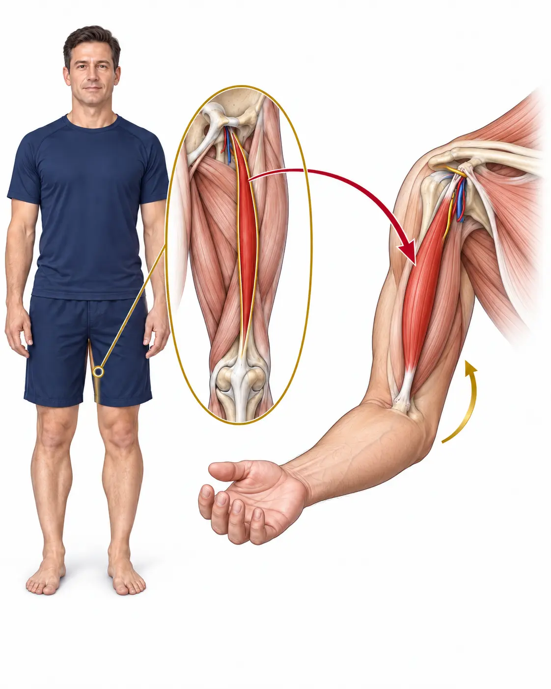
If sufficient spontaneous recovery is unlikely, options may include neurolysis, direct repair, nerve grafting or transferring a functioning donor nerve to a denervated target. Priorities are function-led—for example shoulder stability, elbow flexion and hand function.
Why does timing matter?
Denervated muscle has a limited window for effective neural reconstruction. Significant injuries merit early specialist assessment. Chronic cases may require tendon transfer or free functional muscle transfer.
Upper-extremity nerve entrapment
Numbness, electric sensations, night pain or weakness do not automatically identify the compression site. Cervical root, brachial plexus and more than one point along the same nerve may need consideration.
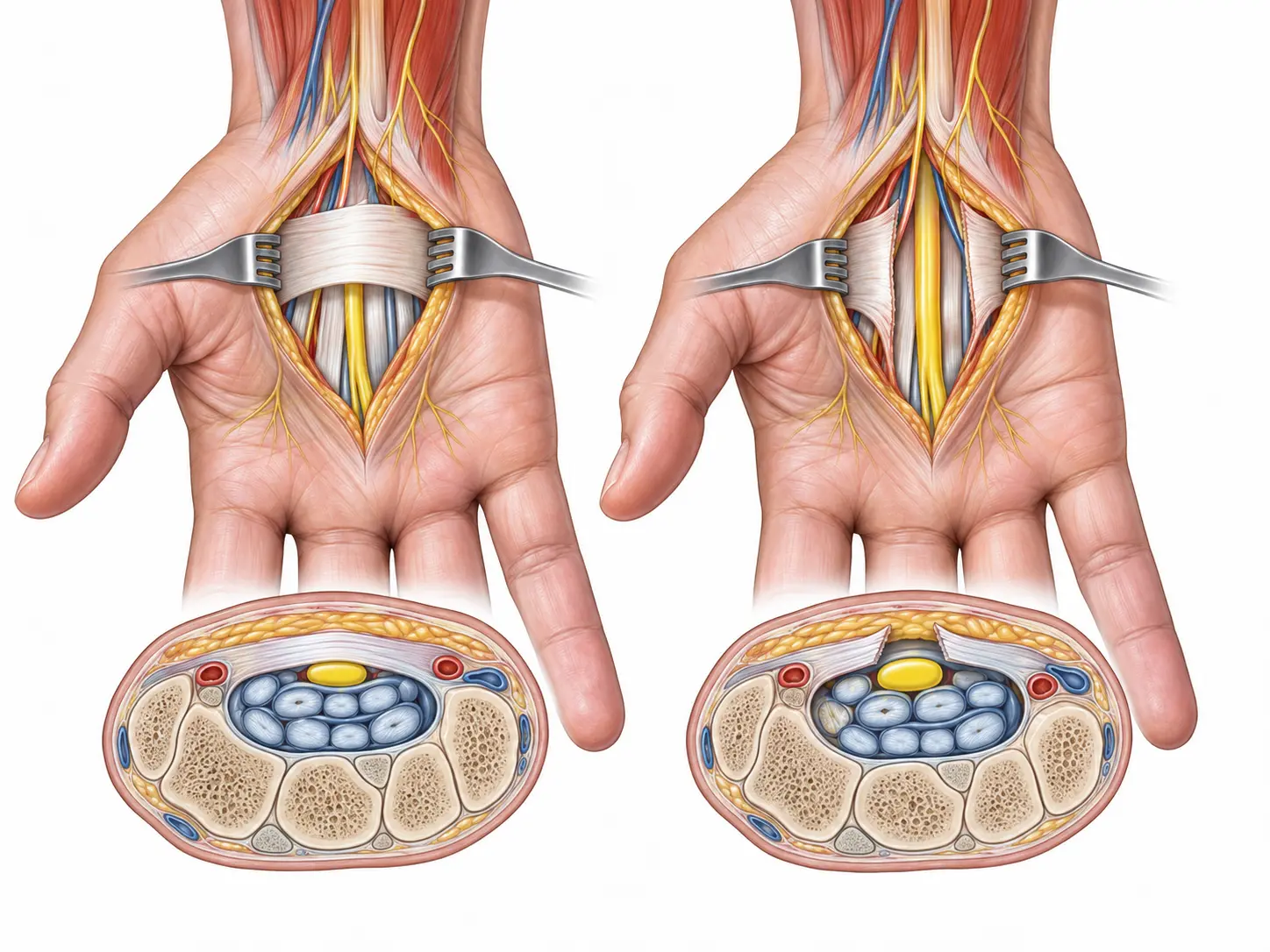
Carpal tunnel syndromeMedian nerve
Median nerve compression at the wrist commonly causes numbness or tingling in the thumb, index, middle and radial half of the ring finger, often at night. Later findings may include pinch weakness or dropping objects; the little finger is usually spared.
Assessment and treatment
Diagnosis is clinical, with nerve studies/EMG or ultrasound used when appropriate. Night splinting, load modification, injection or carpal tunnel release may be considered according to severity, weakness and evidence of nerve injury.
Cubital tunnel syndromeUlnar nerve at the elbow
Ulnar nerve compression at the elbow may cause tingling in the little and ring fingers, elbow-region pain, and weakness of finger spread, key pinch or grip. Prolonged elbow flexion may aggravate symptoms.
Assessment includes sensation, intrinsic strength, nerve stability and alternate compression sites. Initial care may include positional changes and avoiding sustained night flexion; progressive weakness or advanced compression may require decompression and, selectively, anterior transposition.
Guyon canal syndromeUlnar nerve at the wrist
Ulnar nerve compression at the wrist can produce motor, sensory or mixed patterns depending on the zone. Preserved dorsal-ulnar hand sensation can support a distal site but does not establish the diagnosis alone.
Potential causes include a ganglion, repetitive pressure, hamate-region injury or a vascular lesion. Focused examination and selected EMG, ultrasound or MRI guide treatment of both the nerve and the cause of compression.
Radial tunnel and posterior interosseous nerveRadial tunnel · PIN
Deep lateral forearm pain, usually several centimetres below the elbow, may fit radial tunnel irritation. True weakness of finger or thumb extension raises concern for posterior interosseous motor-branch dysfunction and requires precise localization.
Assessment distinguishes a neural source from lateral epicondylitis, cervical disease and tendon pathology. EMG may be normal in pain without weakness; it is more useful when motor loss is present. Care begins with load modification and rehabilitation, with decompression reserved for persistent, well-localized cases.
Cold, colour-changing hand — when it is not a nerveRaynaud and vascular warning
Fingers that turn white or blue in the cold may reflect Raynaud phenomenon, primarily a vascular response rather than a “trapped nerve.” Assessment considers symmetry, triggers and associated disease.
Lower-limb and pelvic neuropathies
Precise localization of pain, numbness or sensitivity helps distinguish a peripheral nerve from plexus, root, joint or soft-tissue pathology. This rubric focuses on sensory nerves of the thigh, knee and pelvis; back pain, foot pain and nerve transfers have their own rubrics.
Lateral femoral cutaneous nerve (LFCN)Meralgia paresthetica
Burning, numbness or hypersensitivity over the outer thigh without muscle weakness may reflect LFCN entrapment. Assessment separates a local nerve problem from spinal, hip or pelvic causes.
Saphenous nerve entrapmentSaphenous nerve
Burning pain or tenderness along the medial knee and leg may follow surgery, trauma or compression in the adductor canal. As this is a sensory nerve, muscle weakness is not typical.
Pudendal neuralgiaPudendal nerve
Perineal pain aggravated by sitting requires a broad differential diagnosis. Care may include seating modification, pelvic-floor therapy, medication and image-guided blocks; surgery is reserved for selected cases.
Facial palsy and smile reconstruction
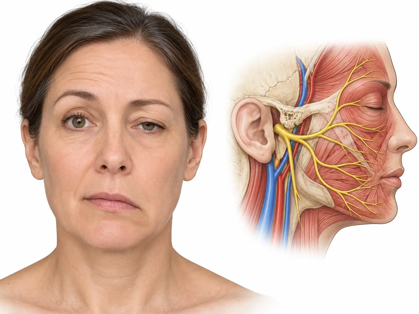
Facial palsy is a sign, not a single diagnosis. Management depends on cause, duration, nerve continuity, remaining muscle function and the presence of synkinesis.
Bell’s palsyAcute facial palsy
Sudden unilateral weakness may cause mouth-corner droop, difficulty smiling or closing the eye, altered tearing or taste, and sometimes pain around the ear.
Facial reanimation and synkinesisDynamic reconstruction
If recovery remains incomplete, care may include specialist facial therapy, botulinum toxin for synkinesis and tailored surgery.
Options include timely nerve repair or grafting, nerve transfers, cross-face nerve grafting, free functional muscle transfer and static support. A recent palsy and longstanding paralysis require different strategies.
Thoracic outlet syndrome
The thoracic outlet is the passage between the clavicle and first rib containing the brachial plexus and major vessels. Symptoms differ according to whether a nerve, vein or artery is compressed.
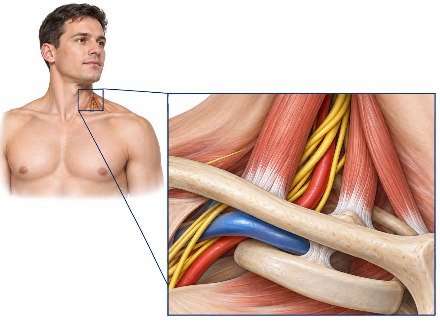
Neurogenic TOSNerve compression
Neck, shoulder, arm or hand pain; tingling—sometimes involving the ring and little fingers—arm fatigue and weakness may occur. Overhead activity can aggravate symptoms, but no single manoeuvre establishes the diagnosis.
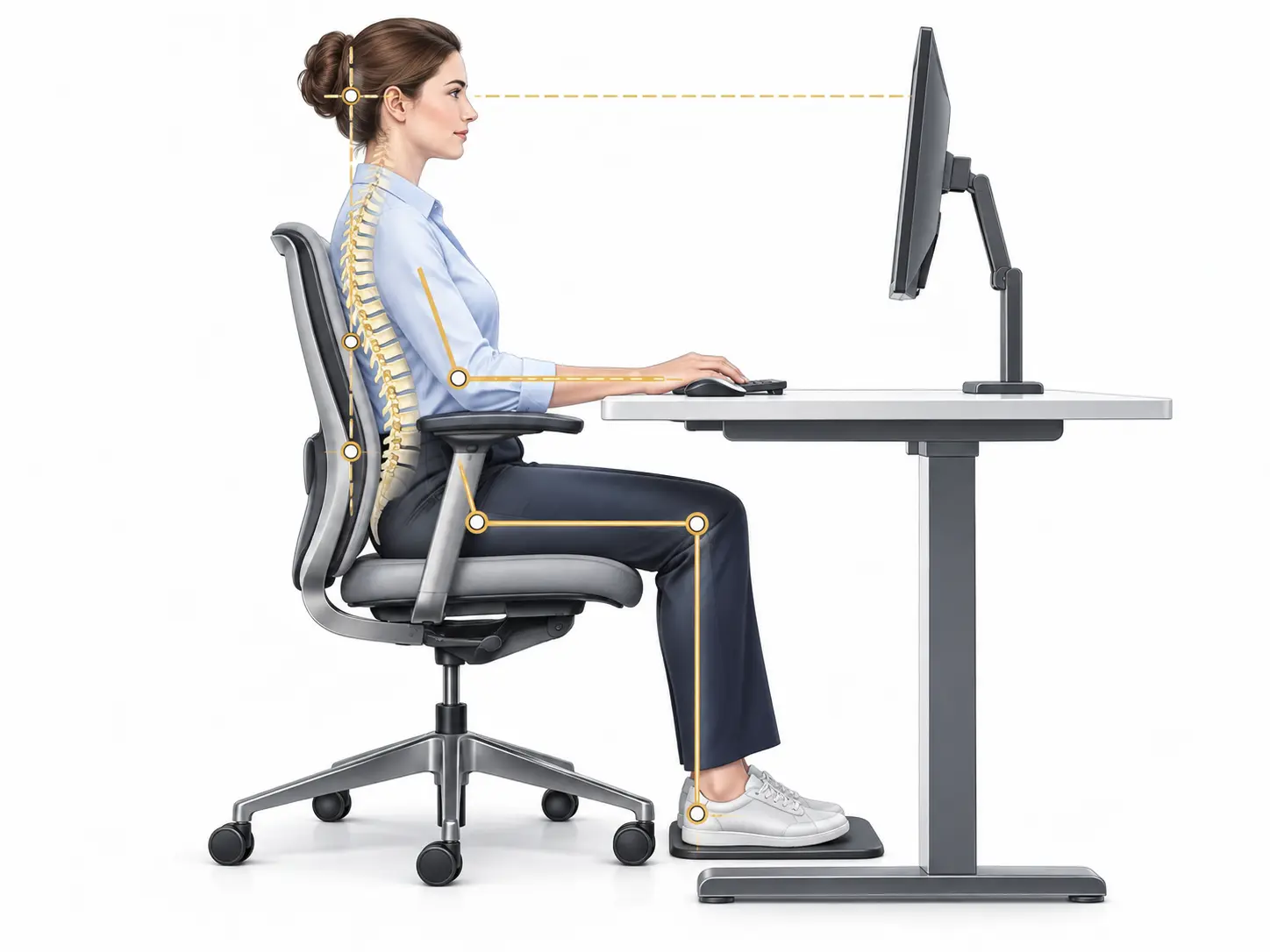
Assessment combines the history, neurologic and mechanical examination, and differentiation from cervical, shoulder and distal nerve disorders. TOS-specific physiotherapy is commonly first-line.
Venous / arterial TOSVascular compression
Sudden swelling, heaviness or blue discoloration may indicate venous involvement. A cold or pale hand, reduced pulse, ischemic pain or fingertip ulceration may indicate arterial compromise.
Treatment and surgeryAnatomy-led
Management is not identical for every patient. Neurogenic TOS usually starts with rehabilitation; surgery is considered when the diagnosis is coherent and substantial symptoms persist despite appropriate care.
Decompression may include scalene, brachial plexus and pectoralis minor release. First-rib resection is not automatic; anatomy, subtype and findings guide the operation.
Migraine, headache and occipital nerves
Migraine is a neurologic disease. Focal peripheral nerve tenderness may coexist in selected patients, but it does not by itself establish the pain source or surgical candidacy.
Treatment before surgical decisionsStaged and multidisciplinary
Management is individualized to the diagnosis and attack burden and may include acute and preventive medication, trigger management and coordinated headache-neurology care.
When indicated, options may include CGRP-targeted biologic therapy, onabotulinumtoxinA (Botox) for chronic migraine, and diagnostic or therapeutic nerve blocks. Surgery is considered only after structured diagnosis, appropriate nonsurgical treatment and careful selection.
Occipital neuralgiaGreater and lesser occipital nerves
Sharp, shooting or stabbing posterior scalp pain, often with focal tenderness and upward radiation, may fit occipital neuralgia. Migraine, cervicogenic headache and other causes must be distinguished.
Greater occipital nerve
Arising mainly from C2, it travels from the occiput toward the posterior and superior scalp. Near-midline tenderness with upward radiation can follow its course.
Lesser occipital nerve
Usually arising from the C2–C3 cervical plexus, it travels along the lateral occiput behind the ear. Lateral occipital or postauricular pain may follow this pathway.
Assessment and treatmentNot every occipital pain is nerve entrapment
Assessment combines the pain pattern, tenderness map, cervical examination and neurologic evaluation. Temporary improvement after local anaesthetic block may support the diagnosis but is not sufficient alone.
Options may include medication, physiotherapy for a cervical component, blocks and injections. Nerve decompression or another operation is reserved for persistent, well-localized pain after appropriate investigation and conservative care.
Breast sensation restoration and pain after breast surgery
After mastectomy or breast reconstruction, two separate goals may be assessed: restoration of meaningful protective sensation and treatment of persistent neuropathic pain after surgery.
Back pain and sciatica from peripheral nerve causes
Some back and buttock pain arises from peripheral nerves outside the spine. Assessment distinguishes root disease from cluneal nerve entrapment or sciatic irritation in the deep gluteal space.
Cluneal nerves and low-back pain
Focal pain over the iliac crest radiating into the buttock or lower back.
Full guide02Deep gluteal and piriformis syndromes
Buttock pain aggravated by sitting and potentially radiating along the sciatic nerve.
Full guideIntercostal neuralgia and chest-wall painIntercostal nerve
Burning, stabbing or scar-line tenderness along a rib can follow intercostal nerve injury after trauma or surgery, but cardiac, pulmonary, spinal and visceral causes must first be excluded. When pain localizes to a nerve, assessment may include mapping, imaging and a diagnostic block; surgery is reserved for selected cases.
Complex procedures performed by Dr Khoury
Selected clinical examples of complex nerve reconstruction, neurolysis, nerve transfer and functional reconstruction. Images on the page come from Dr Khoury’s clinical work and are presented for education.
Lower-extremity nerve transfers
Motor and sensory reconstruction options for foot drop, knee extension and other functions. The page covers all regions in the supplied correct paper: deep peroneal, femoral, sciatic/tibial, obturator and sensory transfers.
Restless legs syndrome
An urge to move the legs, mainly at rest and in the evening, may fit restless legs syndrome. Assessment considers iron deficiency, medication, sleep and other neurologic causes before treatment.
Ankle and foot nerve pain
Pain, numbness, weakness or a “pebble in the shoe” sensation may arise from different nerves. Location and function distinguish common peroneal neuropathy, tarsal tunnel/Baxter nerve and Morton neuroma.
Common peroneal nerveFibular head entrapment
Compression near the fibular head may cause numbness over the dorsum of the foot and weakness lifting the ankle or toes. New foot drop requires early assessment.
Tarsal tunnel and Baxter nerveTibial nerve and plantar branches
Burning or numbness in the sole may reflect compression of the tibial nerve or one of its branches. Assessment also considers spinal disease, neuropathy and local foot pathology.
Morton neuromaInterdigital nerve pain
Burning forefoot pain radiating into the toes, sometimes described as a pebble in the shoe, is often aggravated by narrow footwear and forefoot loading. Treatment begins with footwear modification and progresses according to findings.
Rehabilitation and physiotherapy protocols
Rehabilitation after nerve surgery is staged around tissue protection, range preservation, recognition of reinnervation and motor relearning. Individual instructions from the surgeon and treating therapist always take priority.
This information is general and does not replace diagnosis. Treatment depends on cause, severity, timing, investigations and functional goals. Illustrations are explanatory rather than operative diagrams.
Next step
Focused assessment begins with accurate localization.
Useful records may include previous treatment and operative notes, EMG/nerve-conduction studies, relevant imaging and a list of treatments already attempted.