Planning around vital nerves and vessels
These operations may remove compression, repair or reroute a nerve, restore movement or rebuild sensation. They require anatomical mapping, an appropriate approach, magnification and systematic assessment of nerve function.
Thoracic outlet syndrome
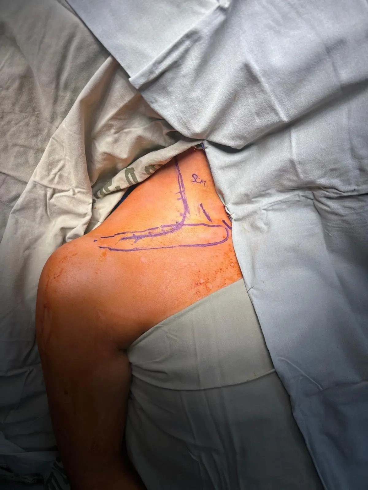
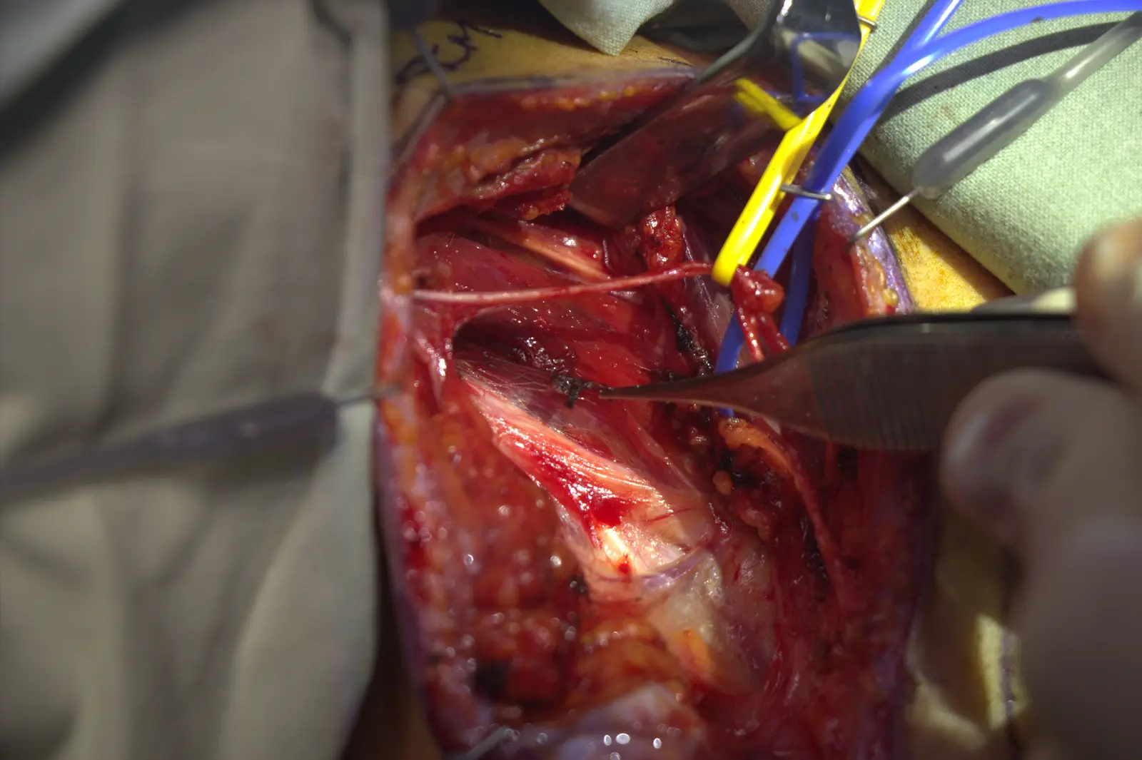
In thoracic outlet syndrome, the brachial plexus or subclavian vessels may be compressed between the neck, first rib and shoulder. Neurogenic, venous and arterial forms require different assessment.
Neurogenic decompression may include scalenectomy, removal of fibrous bands, brachial plexus neurolysis, pectoralis minor release and, when indicated, first- or cervical-rib resection. The operation is not identical for every patient.


Facial movement and smile reconstruction
Planning depends on the duration of paralysis, viability of facial muscles, nerve continuity and available donor nerves. When native muscles remain viable, repair or nerve transfer may be used; longstanding paralysis may require a free functioning muscle such as gracilis, with microsurgical vascular and neural connections.


Nerve decompression
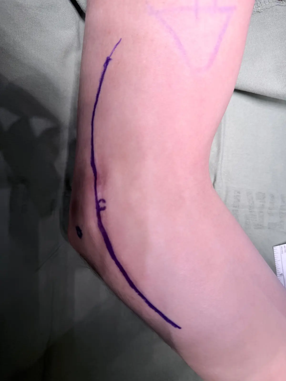
Neurolysis is used when an intact nerve is compressed in a narrow anatomical passage or trapped by scar. The nerve is identified in healthy tissue and followed through the full zone of compression while its blood supply and branches are protected.

Nerve transfers and TMR
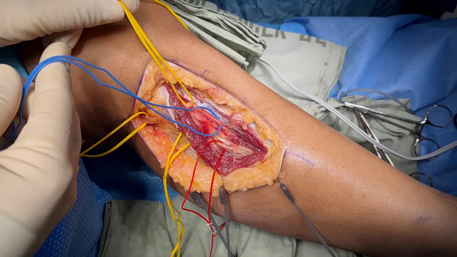
A nerve transfer connects a functioning donor nerve or fascicle to a denervated recipient to bring viable axons closer to a motor or sensory target. Donor cost, timing, muscle viability and axon numbers guide selection.
In targeted muscle reinnervation (TMR), a sensory nerve end is connected to a small expendable motor branch after neuroma management. Complex reconstruction may combine repair, grafting, tendon transfer or free functional muscle transfer.



How the operation is selected
- Define compression, transection, neuroma, motor loss or sensory loss.
- Localize the lesion and determine whether the distal target remains reconstructable.
- Select decompression, direct repair, graft, nerve transfer, TMR or a combined reconstruction.
- Plan rehabilitation and follow-up around nerve-regeneration time and the restored action.
Technical complexity is never an indication by itself. Surgery needs a clear functional or therapeutic goal and a reasonable balance of potential benefit and risk.
Selected references
- Illig KA, Donahue D, Duncan A, et al. Reporting standards of the Society for Vascular Surgery for thoracic outlet syndrome. Journal of Vascular Surgery. 2016;64(3):e23–e35. doi:10.1016/j.jvs.2016.04.039
- Borschel GH, Kawamura DH, Kasukurthi R, et al. The motor nerve to the masseter muscle. JPRAS. 2012;65(3):363–366. doi:10.1016/j.bjps.2011.09.026
- Ray WZ, Mackinnon SE. Management of nerve gaps. Experimental Neurology. 2010;223(1):77–85. doi:10.1016/j.expneurol.2009.03.031