Anatomy and the three subtypes
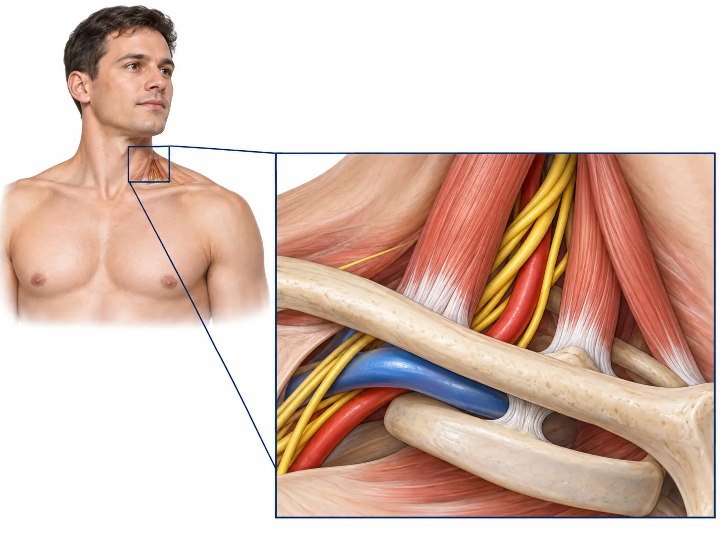
Neurogenic TOS involves the brachial plexus and is the most common subtype. Venous TOS affects the subclavian vein and may be associated with thrombosis. Arterial TOS affects arterial flow and may threaten the hand. These are not interchangeable diagnoses and do not follow the same pathway.
Symptoms and red flags
- Neurogenic: neck, shoulder, arm or hand pain, tingling, fatigue and weakness, sometimes aggravated by overhead activity.
- Venous: sudden swelling, heaviness, blue discoloration or prominent arm veins.
- Arterial: a cold or pale hand, pulse change, ischemic pain or fingertip ulceration.
How TOS is diagnosed
No single test confirms neurogenic TOS in every patient. Diagnosis integrates the symptom pattern, neurologic and mechanical examination, cautious use of provocative manoeuvres, and exclusion of cervical, shoulder, cubital tunnel and carpal tunnel disorders.
Radiographs can identify a cervical rib or bony variation. EMG and nerve-conduction studies are selected according to the presentation, often to identify or exclude another neuropathy. Suspected venous or arterial disease requires Doppler and appropriate vascular imaging. One positive test or manoeuvre is not enough to plan surgery.
Nonoperative care
The INTOS consensus generally supports conservative management first for neurogenic TOS, apart from selected patients with substantial muscle atrophy or weakness. Care may include load modification, breathing and mobility work, scapular control, graded strength and workstation changes. The aim is not forceful “nerve stretching” but improved movement and tolerance without repeatedly provoking symptoms.
When decompression is considered
Surgery may be considered when the diagnosis is coherent, symptoms remain substantial despite appropriate care, or progressive neural injury is present. Venous and arterial TOS follow a vascular pathway and may require more urgent multidisciplinary treatment.
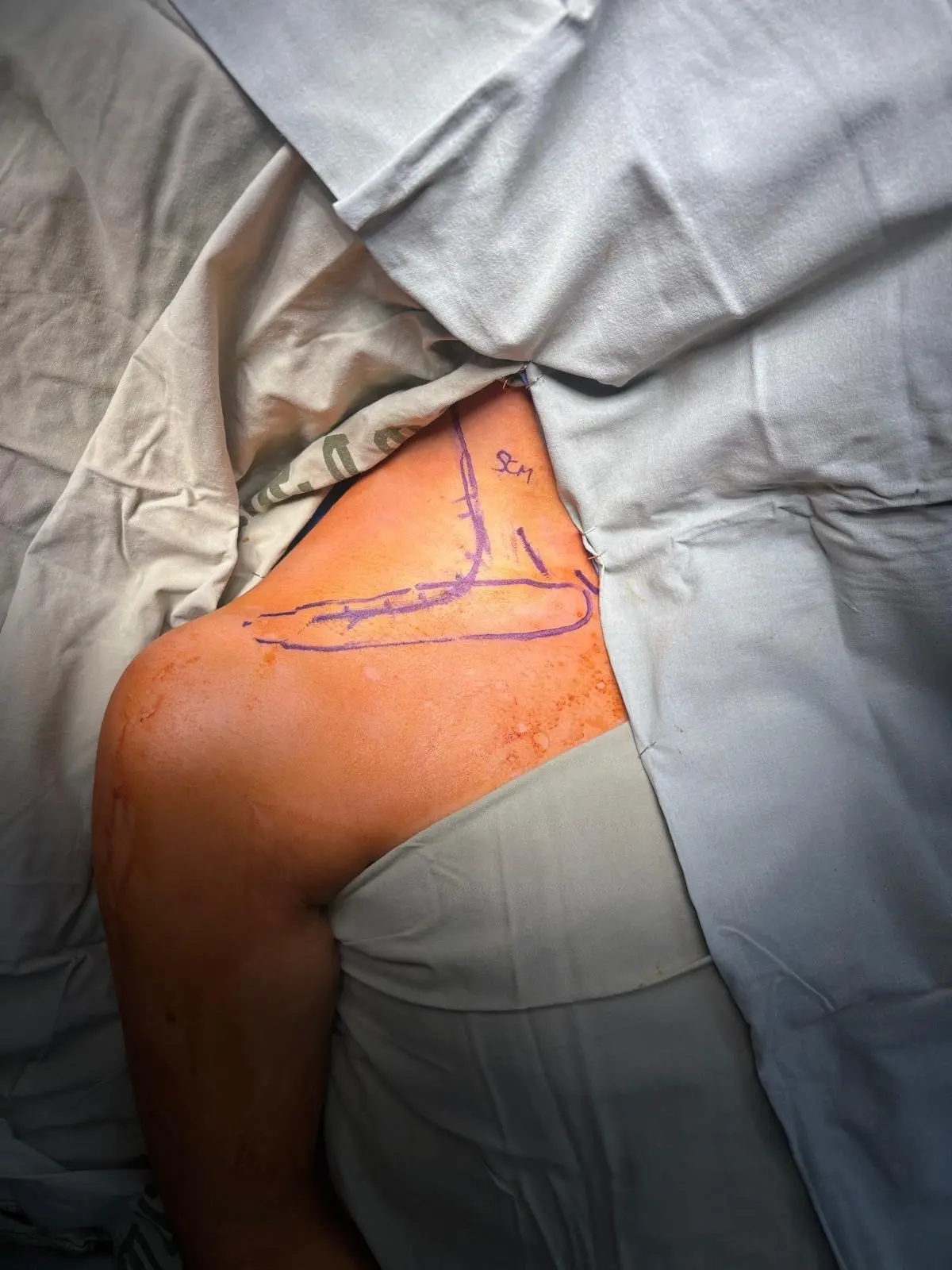
Decompression is matched to the site of compression and may include scalene treatment, brachial plexus release, pectoralis minor release and, in selected cases, first-rib resection. Rib resection is not automatic in every case; subtype, anatomy and findings determine the operation.
Rehabilitation and follow-up
Postoperative rehabilitation is also staged. Early care protects tissues while maintaining comfortable motion; scapular control, endurance, strength and gradual return to work or sport follow. New swelling, discoloration, fever, shortness of breath or neurologic deterioration requires contact with the treating team.
Key references
- Chim H, Hagan RR. Consensus Recommendations for Neurogenic Thoracic Outlet Syndrome from the INTOS Workgroup. Plastic and Reconstructive Surgery – Global Open. 2024;12:e6107. doi:10.1097/GOX.0000000000006107
- Rochkind S, et al. Thoracic Outlet Syndrome Part II: Consensus on the Management of Neurogenic Thoracic Outlet Syndrome by the EANS Section of Peripheral Nerve Surgery. Neurosurgery. 2023;92(2):251–257. doi:10.1227/neu.0000000000002232