Typical symptoms
The ulnar nerve passes behind the inner side of the elbow—the “funny bone” region. Ongoing compression can cause tingling in the little finger and ulnar half of the ring finger, elbow or forearm pain, grip weakness and difficulty with fine tasks.
More advanced injury can produce intrinsic hand-muscle wasting, reduced finger spreading, weak key pinch and altered finger posture. Sensory symptoms alone do not always mean mild disease; examination of muscle function and nerve studies matters.
Why compression occurs
Elbow flexion stretches the nerve and reduces tunnel volume. Sleeping with a flexed elbow, prolonged leaning, repetitive load, scar, bony change or nerve instability may contribute. The nerve can move over the medial epicondyle during flexion; this influences planning but does not by itself prove the source of symptoms.
Diagnosis and nerve severity
Examination maps sensation, intrinsic hand strength, pinch and grip, nerve tenderness and stability during flexion. The neck, shoulder and wrist are assessed to identify an alternate source or more than one compression site.
Nerve-conduction studies and EMG can localize slowing and estimate axonal loss; ultrasound can demonstrate enlargement, compression and movement of the nerve. A normal study does not invariably exclude early or dynamic disease, so results are interpreted with the clinical pattern.
Nonoperative care
When there is no progressive weakness or advanced nerve injury, care may begin with avoiding direct pressure and sustained flexion, modifying sleep position and sometimes using a soft night support. Workstation adjustment and guided exercise can reduce irritation. Nerve-gliding exercises should be gentle and stopped if they produce persistent tingling or pain.
Surgical options
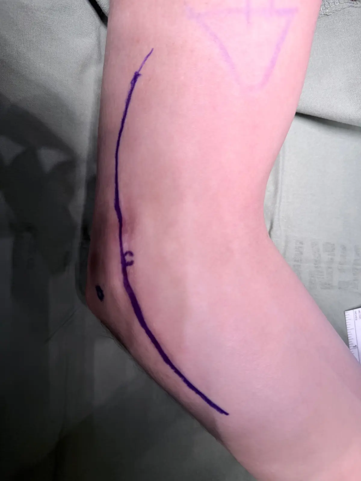
Surgery may be considered for weakness or axonal injury, moderate-to-severe compression, or symptoms that remain limiting despite suitable care. Options include open in-situ release, endoscopic release, and subcutaneous or submuscular anterior transposition. A 2024 review of randomized trials did not find a statistically significant difference in treatment response among the four techniques studied, while complications and reported outcomes varied. Technique is therefore selected around nerve stability, anatomy and the clinical problem.
With severe axonal loss, a distal motor nerve transfer may be considered as an adjunct in selected patients to provide a closer neural source for intrinsic hand muscles. It does not automatically replace decompression of the primary compression site.
Recovery and expectations
Intermittent tingling may improve earlier, while a chronically compressed nerve with axonal loss recovers slowly. Strength recovery depends on injury severity, duration and distance to target muscles. Surgery first aims to stop continued compression; function already lost cannot always be restored completely.
Key reference
- Abourisha E, Srinivasan AS, Barakat A, Chong HH, Singh HP. Surgical management of cubital tunnel syndrome: a systematic review and meta-analysis of randomised trials. Journal of Orthopaedics. 2024;53:41–48. doi:10.1016/j.jor.2024.02.041