Typical symptoms
The common peroneal nerve curves around the fibular neck at the outer knee and divides into deep and superficial branches. Injury may cause weak ankle and toe dorsiflexion, weak eversion, a steppage gait and altered sensation over the lateral leg and dorsum of the foot.
Possible causes
Compression near the fibular head can follow prolonged leg crossing or squatting, rapid weight loss, pressure from a cast or dressing, prolonged immobility, knee-region surgery, or a mass such as a ganglion. Trauma may stretch or rupture the nerve. Diabetes and polyneuropathy may increase susceptibility but do not explain every focal weakness.
Not every foot drop is peroneal neuropathy
Dorsiflexion weakness may also arise from the L5 root, sciatic nerve, lumbosacral plexus, a central neurologic disorder or muscle disease. Sensory mapping and testing foot inversion and proximal muscles help localization but do not replace a complete assessment.
Diagnosis and monitoring
Examination maps strength, sensation, gait and tenderness around the fibular head. EMG and nerve-conduction studies help localize the lesion, estimate axonal loss and identify signs of recovery. Ultrasound or MRI may show compression, internal nerve change, a ganglion or structural injury. Imaging is selected around the injury mechanism and clinical findings.
Nonoperative treatment and rehabilitation
When the pattern suggests reversible pressure, care begins by removing the cause, avoiding fibular-head pressure, preserving ankle and knee motion, training remaining active muscles and improving gait safety. An ankle-foot orthosis can reduce tripping and protect the ankle during recovery. Areas with sensory loss must be protected, and the brace should be checked so it does not create new pressure.
Decompression, repair and nerve transfer
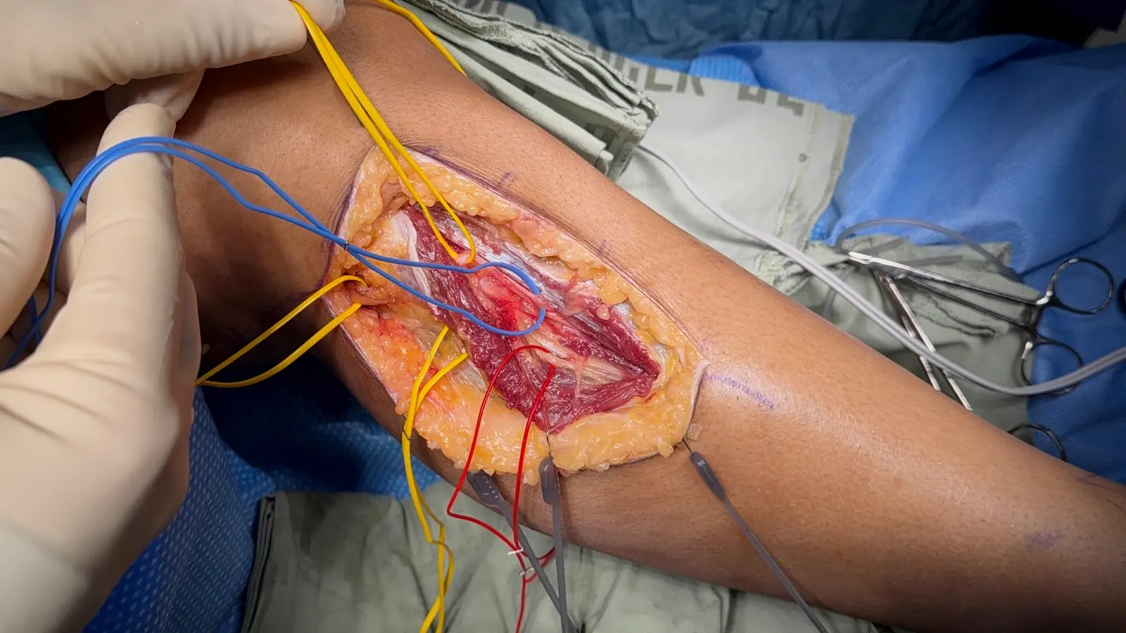
Decompression may be considered when findings localize pressure, weakness or pain persists, a structural cause is present, or recovery is insufficient. Open injury or rupture may require repair or grafting. In selected cases and a suitable time window, nerve transfer may be considered; when muscle is no longer reinnervatable, tendon transfer provides another functional option.
The decompression literature is largely composed of non-uniform series. A meta-analysis reported improved dorsiflexion strength after common peroneal neurolysis while emphasizing the need for higher-quality studies. Individual decisions should not be based on a single success percentage.
Key references
- Wilson C, Yaacoub AP, Bakare A, et al. Peroneal nerve decompression: institutional review and meta-analysis to identify prognostic associations with favorable and unfavorable surgical outcomes. Journal of Neurosurgery: Spine. 2019;30(5):714–721. doi:10.3171/2018.10.SPINE18626
- Chow AL, Levidy MF, Luthringer M, Vasoya D, Ignatiuk A. Clinical Outcomes After Neurolysis for the Treatment of Peroneal Nerve Palsy: A Systematic Review and Meta-Analysis. Annals of Plastic Surgery. 2021;87(3):316–323. doi:10.1097/SAP.0000000000002833