Symptoms and red flags
The presentation can include burning or electric pain, tingling, sensory loss and weakness involving the shoulder, elbow, wrist or fingers. A partial lesion preserves some functions; a more extensive injury can leave the arm flaccid and insensate.
Injury patterns: stretch, rupture or avulsion
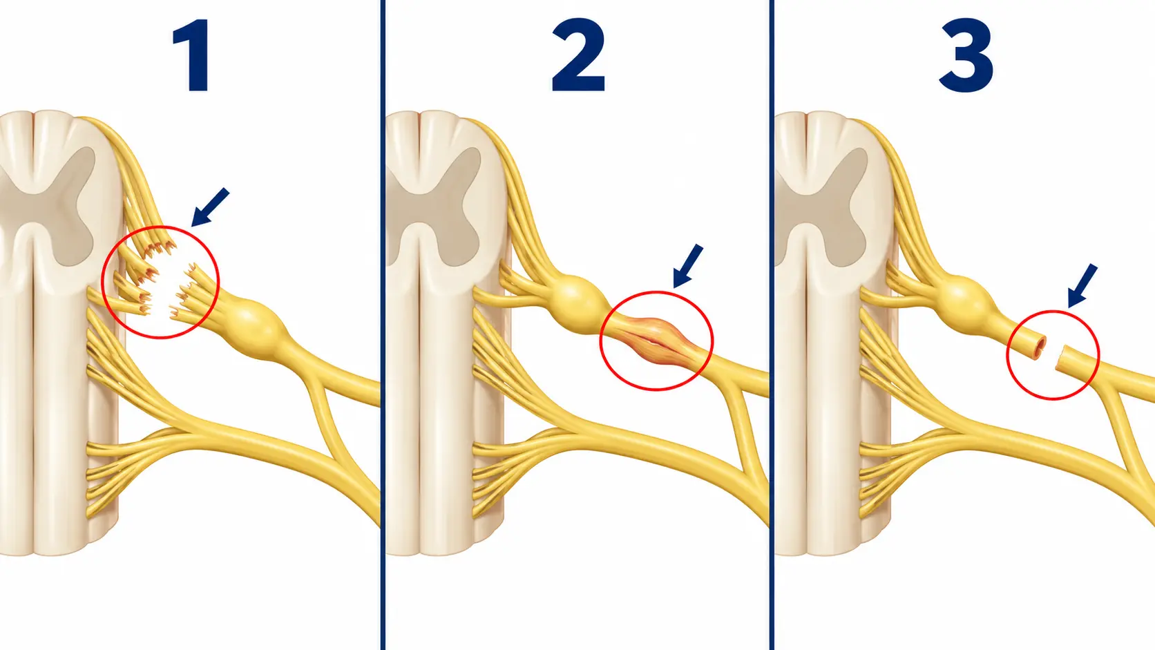
With a lesser stretch injury, nerve continuity may be preserved and recovery may occur. A rupture disrupts the nerve outside the spinal cord. An avulsion separates the nerve root from the cord and cannot be repaired in the same way. Mixed levels and injury types may coexist in one limb.
Assessment and active monitoring
Assessment begins with the injury mechanism and systematic mapping of strength, sensation, pain and joint motion. EMG and nerve-conduction studies, MRI or MR neurography, and sometimes CT myelography are selected according to the question and timing.
A single test is not always decisive. Serial examination can show whether reinnervation and recovery are occurring. During this period, therapy protects joint motion, limits contracture and helps safeguard areas with sensory loss.
Why timing matters
Not every injury requires surgery, but observation should not be open-ended. After denervation, muscle progressively loses its capacity to accept reinnervation. A systematic review of traumatic stretch and blunt injuries generally found better motor outcomes when required reconstruction occurred within six months, while emphasizing the balance with possible spontaneous recovery.
This is not an automatic deadline for every lesion. Laceration, avulsion, childhood injury and clear recovery patterns require different decisions. Early specialist referral allows a monitoring plan without inadvertently losing a possible reconstructive window.
Surgical options
Neurolysis and repair
Releasing a scarred nerve or directly repairing it when nerve ends and tension permit.
Nerve graft
Bridging an injured segment so axons can grow toward an appropriate target.
Nerve transfer
Connecting an expendable functioning branch to a nearby target nerve to shorten the route to muscle.
Functional muscle transfer
Transferring muscle with its vessels and nerve when the original muscle is no longer reconstructible.
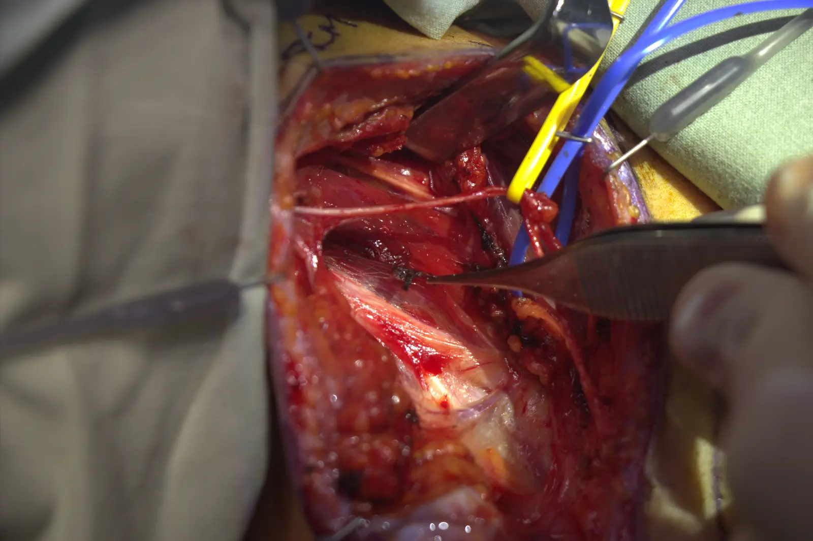
Planning prioritizes meaningful goals such as shoulder stability, elbow flexion, protective sensation and useful hand function. Complete recovery cannot be guaranteed, and more than one reconstructive technique may be combined.
Rehabilitation and expectations
Rehabilitation starts with joint protection and preservation of motion. As reinnervation appears, motor relearning is introduced: initially activating the donor movement and then learning to recruit the new target more independently. Recovery unfolds over months and requires coordinated surgical and therapy follow-up.
Key reference
- Martin E, Senders JT, DiRisio AC, Smith TR, Broekman MLD. Timing of surgery in traumatic brachial plexus injury: a systematic review. Journal of Neurosurgery. 2019;130(4):1333–1345. doi:10.3171/2018.1.JNS172068